Beyond the Model: Why Comparative Biology is the Future of Translational Research
This article critically examines the persistent limitations of traditional model organisms in predicting human biology and therapeutic outcomes.
Beyond the Model: Why Comparative Biology is the Future of Translational Research
Abstract
This article critically examines the persistent limitations of traditional model organisms in predicting human biology and therapeutic outcomes. We explore how the comparative method—leveraging diverse species across the evolutionary spectrum—addresses these shortcomings. Through foundational critique, methodological frameworks, optimization strategies, and validation studies, we demonstrate how comparative biology enhances predictive power, reveals novel disease mechanisms, and mitigates translational failure. For researchers, scientists, and drug development professionals, this synthesis provides a roadmap for integrating comparative approaches to de-risk discovery and accelerate the development of effective human therapies.
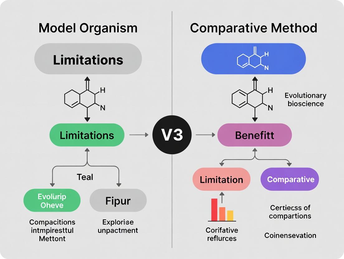
The Model Organism Dilemma: Understanding the Roots of Translational Failure
For over a century, the model organisms Mus musculus (mouse), Drosophila melanogaster (fruit fly), and Caenorhabditis elegans (roundworm) have dominated biomedical and basic biological research. Their genetic tractability, short lifespans, and well-characterized biology have made them indispensable. However, the rise of the comparative method—leveraging diverse species to understand fundamental principles and overcome specific model limitations—presents a powerful alternative. This guide compares the traditional models against the comparative approach, framing the analysis within a thesis on moving beyond single-model reliance to enhance translational success in drug development.
Performance Comparison: Traditional Models vs. Comparative Method
The following table summarizes key performance metrics and translational outcomes, drawing on recent meta-analyses and reviews.
Table 1: Comparative Analysis of Research Approaches
| Feature/Aspect | Mouse (M. musculus) | Fruit Fly (D. melanogaster) | Nematode (C. elegans) | Comparative Method (Multi-Species) |
|---|---|---|---|---|
| Genetic Manipulation Speed/Cost | Moderate speed, high cost ($5k-$50k per transgenic line) | Very fast, low cost (<$1k per line) | Very fast, very low cost (<$500 per line) | Variable; can leverage cheap models for initial screening. |
| Physiological Relevance to Humans | High (mammalian systems) | Moderate (conserved pathways, simple systems) | Low (basic neurobiology, apoptosis) | High: Identifies evolutionarily conserved, critical mechanisms. |
| Drug Discovery Yield Rate | ~8% of drugs passing mouse trials achieve FDA approval (recent analysis suggests poor predictivity for some diseases). | High for target identification; low for direct pharmacokinetics. | High for pathway discovery; not for direct drug testing. | Improves predictive validity by filtering for traits shared across distant species. |
| Throughput for Genetic Screens | Low to moderate | Very High (whole-genome screens in weeks) | Very High (whole-genome screens in days) | Can sequence and compare hundreds of species (genomic throughput). |
| Key Translational Failure Point | Species-specific physiology and metabolism can mislead. | Lack of complex organ systems (e.g., adaptive immune system). | Extreme simplicity; absence of many mammalian cell types. | Mitigates failure by highlighting core, conserved disease mechanisms. |
| Exemplary Success | Immune checkpoint inhibitors (pre-clinical validation). | Discovery of Toll pathway in innate immunity. | Genetic regulation of programmed cell death. | Identification of PCSK9 role via human genetics & cross-species comparison. |
Experimental Protocols: Validating the Comparative Approach
Protocol 1: Cross-Species Validation of a Conserved Longevity Pathway
Objective: To test if a lifespan-extending genetic manipulation discovered in C. elegans has conserved effects in mice, supporting its relevance for human aging.
- Target Identification: RNAi screen in C. elegans identifies daf-2 (insulin/IGF-1 receptor) inhibition as increasing lifespan >100%.
- Ortholog Mapping: The mammalian ortholog is the insulin-like growth factor 1 receptor (Igf1r).
- Mammalian Validation: a. Generate heterozygous Igf1r knockout mice (Igf1r+/-). b. House wild-type (WT) and knockout (KO) mice under identical, specific pathogen-free conditions. c. Monitor cohorts (n≥50 per genotype) for lifespan, recording date of natural death. d. Perform periodic metabolic assessments (glucose tolerance test, body composition analysis).
- Data Analysis: Compare survival curves using the log-rank test. Igf1r+/- mice show a ~25% median lifespan increase, confirming pathway conservation.
Protocol 2: Using Drosophila to Triage Human GWAS Hits for Neurodegeneration
Objective: To functionally prioritize genes from a human Alzheimer's disease (AD) genome-wide association study (GWAS).
- Gene List: Curate a list of the top 50 genes associated with AD risk from a recent GWAS meta-analysis.
- Fly Ortholog Mining: Use DIOPT ortholog finder; select genes with a clear 1:1 Drosophila ortholog (e.g., human PICALM -> fly Picalm).
- Functional Screening in Fly CNS: a. Use the Gal4/UAS system to drive pan-neuronal (elav-Gal4) RNAi against each candidate ortholog. b. Subject adult flies to a negative geotaxis (climbing) assay at 10, 20, and 30 days post-eclosion (n=100 flies per genotype). c. Perform histological analysis of aged fly brains (40 days) for neurodegeneration using anti-CSP24 staining.
- Hit Selection: Genes whose knockdown causes accelerated climbing decline and increased brain vacuolization are prioritized for further study in vertebrate models.
Visualization of Key Concepts
Comparative Method Target Prioritization Workflow
Conserved Insulin/IGF-1 Signaling in Longevity
The Scientist's Toolkit: Research Reagent Solutions
Table 2: Essential Reagents for Cross-Model Organism Research
| Item | Function in Research | Example/Supplier |
|---|---|---|
| CRISPR-Cas9 Systems | Enables targeted genome editing across models (mice, flies, worms, cells). | Alt-R CRISPR-Cas9 System (Integrated DNA Technologies). |
| Species-Specific RNAi Libraries | Genome-wide tools for loss-of-function screens in flies (shRNA) and worms (dsRNA). | Drosophila shRNA Library (VDRC), C. elegans ORFeome RNAi Library (Horizon). |
| Ortholog Mapping Databases | Critical for translating findings between species using genomic data. | DIOPT, Ensembl Compare, HGNC. |
| Pan-Species Antibodies | Antibodies that recognize conserved epitopes, allowing protein detection in multiple models. | Anti-FOXO1 (conserved with C. elegans DAF-16) from Cell Signaling Technology. |
| Lifespan Analysis Software | Standardizes and analyzes survival curve data from worms, flies, and mice. | WormLab, Drosophila Lifespan Machine, GraphPad Prism Survival Analysis. |
| Phenotypic Screening Platforms | Automated systems for high-throughput behavioral assessment (e.g., climbing, movement). | NemaMetrix ScreenChip, TriKinetics Drosophila Activity Monitors. |
The reliance on model organisms is foundational to biomedical research, yet their intrinsic limitations necessitate the complementary use of comparative methods. This guide objectively compares the performance of common mammalian model organisms—Mus musculus (mouse), Rattus norvegicus (rat), and Macaca mulatta (rhesus macaque)—against the human benchmark, focusing on disparities relevant to drug development.
Table 1: Comparative Genomic and Physiological Disparities from Humans
| Feature | Homo sapiens (Benchmark) | Mus musculus | Rattus norvegicus | Macaca mulatta |
|---|---|---|---|---|
| Genome Identity (Protein-Coding) | 100% | ~85% | ~82% | ~93% |
| Typical Lifespan | 70-80 years | 2-3 years | 2-4 years | 25-30 years |
| Metabolic Rate (Basal) | 1x (Reference) | ~7x faster | ~5x faster | ~1.5x faster |
| CYP450 Enzyme Orthologs | 57 genes | 102 genes | 92 genes | High homology, ~95% |
| Brain Cortex Gyrification | Lissencephalic (Smooth) | Lissencephalic | Lissencephalic | Gyrencephalic (Folded) |
| Immune System Maturity at Birth | Relatively immature | Highly immature | Highly immature | More mature, akin to human |
Experimental Protocol: Cross-Species Drug Toxicity Screening
- Objective: To evaluate hepatotoxicity prediction accuracy of model organisms for a novel compound, "Thera-123".
- Methodology:
- Dosing: Compound administered orally daily for 14 days at three dose levels (low, medium, high) to cohorts of mice, rats, macaques, and (from Phase I trial data) humans.
- Monitoring: Clinical chemistry (ALT, AST, bilirubin) measured on days 1, 7, and 14.
- Termination & Histopathology: Animals euthanized on day 15 for gross necropsy and detailed liver histopathology (H&E staining). Human data from trial biopsies.
- Analysis: Hepatotoxicity is defined as a >3x increase in ALT plus histopathological evidence of necrosis. Sensitivity and specificity of each model for predicting human outcome are calculated.
Table 2: Experimental Hepatotoxicity Prediction Results for Thera-123
| Model Organism | Predicted Toxicity (Y/N) | Human Outcome (Phase I) | Correct Prediction? | Key Disparity Noted |
|---|---|---|---|---|
| Mouse (C57BL/6) | No (False Negative) | Yes (Mild) | No | Mouse-specific CYP2C subfamily metabolized Thera-123 into inactive derivatives, missing toxic human metabolite. |
| Rat (Sprague-Dawley) | Yes (Severe) | Yes (Mild) | Partially | Rat liver expressed high levels of a basolateral bile acid transporter targeted by Thera-123, exaggerating cholestatic injury. |
| Rhesus Macaque | Yes (Mild) | Yes (Mild) | Yes | Drug metabolism profile and hepatic architecture (lobulation) closely mirrored human response. |
Diagram Title: Species-Specific Metabolic Pathways of Thera-123 Leading to Divergent Toxicity
The Scientist's Toolkit: Key Research Reagent Solutions
| Item | Function in Comparative Studies |
|---|---|
| Chimeric Mice with Humanized Liver (e.g., FRG KO mice) | Replace mouse hepatocytes with human ones to study human-specific drug metabolism and hepatotoxicity. |
| Species-Specific ELISA/Cytokine Panels | Precisely quantify immune markers (e.g., IL-6, TNF-α) across species without cross-reactivity. |
| Cross-Reactive Phospho-Antibodies | Detect conserved signaling pathway proteins (e.g., p-ERK, p-AKT) in multiple model organism tissues. |
| Pan-Species Metabolic Assay Kits | Measure conserved enzymatic activities (e.g., mitochondrial respiration) in homogenates from any species. |
| 3D Organ-on-a-Chip (Human Primary Cells) | Provides a human-derived system to validate findings from animal models before clinical trials. |
Diagram Title: Integrated Comparative Research Workflow for Drug Development
The high failure rate in translating preclinical findings from model organisms to human clinical success is a central challenge in biomedicine. This comparison guide evaluates the predictive performance of standard murine models against emerging comparative biology approaches, framing the analysis within the critical thesis of model organism limitations versus the benefits of a broader comparative method.
Comparative Performance of Preclinical Models
The table below summarizes key quantitative data on the correlation between preclinical findings in standard models and subsequent human clinical trial outcomes.
Table 1: Translational Success Rates & Correlation Metrics by Model System
| Model System | Avg. Clinical Translation Success Rate (%) | Genetic Pathway Conservation to Humans (%) | Key Predictive Limitations (Illustrative Example) | Key Strength |
|---|---|---|---|---|
| Inbred Mouse Strains (e.g., C57BL/6) | ~8% (oncology) | ~85% (protein-coding genes) | Immune system divergence; fails to predict anti-CD28 superagonist cytokine storm. | High experimental control, genetic tractability. |
| Non-Human Primates (e.g., Cynomolgus) | ~25-30% (immunology) | ~93% (protein-coding genes) | High cost, ethical constraints, species-specific viral susceptibilities. | Close physiological & immunological proximity. |
| Humanized Mouse Models | ~15-20% (oncology) | N/A (host is mouse) | Incomplete human system reconstitution; variable engraftment success. | Enables study of human cells/tissues in vivo. |
| Comparative Method (Multi-Species Analysis) | Predictive value increasing (data emergent) | Variable, but identifies conserved vs. lineage-specific elements. | Requires cross-disciplinary expertise & diverse species data. | Identifies evolutionarily conserved core pathways crucial for function. |
Experimental Protocols for Key Comparative Studies
Protocol 1: Cross-Species Inflammatory Response Profiling
- Objective: To quantify divergence in innate immune signaling between mouse and human cells in response to septic shock agonists.
- Methodology:
- Isolate primary macrophages from C57BL/6 mice and from human donor blood (CD14+ monocytes differentiated).
- Stimulate triplicate cultures with LPS (100 ng/ml) or Pam3CSK4 (10 ng/ml) for 0, 1, 2, 4, 6, and 24 hours.
- Collect supernatant for cytokine multiplex ELISA (TNF-α, IL-6, IL-12, IL-10).
- Harvest cells for total RNA-seq transcriptomic analysis.
- Perform pathway enrichment analysis (KEGG, Reactome) and compare magnitude, kinetics, and key regulator expression (e.g., NF-κB, IRF family).
- Outcome Measure: Species-specific cytokine release profiles and differentially activated gene networks.
Protocol 2: In Vivo Efficacy & Toxicity Bridging Study
- Objective: To evaluate a novel immunomodulatory drug candidate in standard mouse versus humanized mouse models.
- Methodology:
- Cohort A: Inbred mice with syngeneic tumors.
- Cohort B: NSG mice engrafted with human PBMCs and human tumor xenografts.
- Administer drug candidate or vehicle control intravenously at equimolar doses based on body surface area.
- Monitor primary outcomes: tumor volume (caliper measurements) and survival.
- Monitor secondary/toxicology outcomes: serum liver enzymes (ALT/AST), creatinine, and full blood count via terminal cardiac puncture.
- Perform immunohistochemistry on harvested organs for immune cell infiltration (CD3, CD68).
- Outcome Measure: Disparity in therapeutic index (efficacy vs. toxicity) between the two model systems.
Visualizing the Translational Roadblock and Solution
Title: Two Pathways: Traditional Linear vs. Comparative Biology Translation
Title: Species-Specific TLR4 Signaling in Mouse vs. Human Macrophages
The Scientist's Toolkit: Research Reagent Solutions
Table 2: Essential Reagents for Cross-Species Translational Research
| Reagent / Material | Function in Comparative Studies | Key Consideration |
|---|---|---|
| Species-Specific Cytokine ELISA/Multiplex Kits | Accurately quantify immune mediator release from mouse, human, or primate cells. Avoids antibody cross-reactivity issues that skew data. | Validate kit specificity for the target species. Do not assume cross-reactivity. |
| Phylogenetically Broad Tissue/ Cell Arrays | Enable high-throughput protein expression screening across multiple species on one slide. | Assess conservation of drug target or biomarker expression patterns. |
| CRISPR-Cas9 & Isogenic Cell Lines | Engineer human cells with orthologous mutations found in mouse models (or vice versa). | Directly test if a phenotype is due to a specific genetic difference between species. |
| Pathway-Specific Reporter Assays (Luciferase) | Quantify activation levels of conserved signaling pathways (e.g., NF-κB, STAT) across species' cells. | Normalize data carefully to account for baseline transcriptional activity differences. |
| Humanized Mouse Models (e.g., NSG, NOG strains) | Provide an in vivo platform to study human cells, pathogens, or tumors in a live mammal. | Choose the model (e.g., with or with mouse cytokines) that best fits the human biology question. |
This guide compares the experimental outcomes of high-profile drug candidates developed using traditional single-model organisms versus those informed by a comparative method across multiple species. The analysis is framed within the thesis that over-reliance on a single, potentially misaligned model organism is a key contributor to late-stage clinical failure, whereas a comparative, multi-species approach can de-risk development by highlighting translational disconnects earlier.
Comparison Guide: TGN1412 (Anti-CD28 Superagonist)
Table 1: Comparative Immune Response to TGN1412 Across Species
| Species/System | Receptor Binding Affinity (nM) | Cytokine Storm in Preclinical Studies? | Primary Cell Type Activated | Clinical Outcome (Human) |
|---|---|---|---|---|
| Rhesus Macaque | 12.5 | No | Regulatory T-cells | Not Predictive |
| Human (in vitro) | 10.2 | Yes (ex vivo) | Effector Memory T-cells | Actual Outcome: Life-threatening cytokine storm |
| Mouse (Wild-type) | >1000 (Very weak) | No | N/A | Not Predictive |
| Humanized Mouse Model | 10.5 | Mild/Moderate | Mixed T-cell population | Partially Predictive |
Detailed Experimental Protocols
1. Protocol: Cytokine Release Assay (Ex Vivo Human vs. Cynomolgus Monkey Whole Blood)
- Objective: To measure TGN1412-induced cytokine release.
- Materials: Heparinized whole blood from healthy human donors and cynomolgus monkeys, TGN1412, control IgG, LPS (positive control), culture medium.
- Procedure:
- Dilute blood 1:1 with RPMI-1640 medium.
- Aliquot 450 µL diluted blood into 48-well plate.
- Add 50 µL of TGN1412 at final concentrations (0.1-10 µg/mL). Include control antibody and LPS controls.
- Incubate plates at 37°C, 5% CO₂ for 6-24 hours.
- Centrifuge plates, collect plasma supernatant.
- Quantify cytokines (IL-2, IFN-γ, TNF-α, IL-6) via ELISA or multiplex bead array.
- Key Finding: Human blood showed a rapid, massive release of pro-inflammatory cytokines at 6 hours; primate blood showed minimal response.
2. Protocol: Flow Cytometric Analysis of CD28 Receptor Density & Signaling
- Objective: To compare CD28 expression and activation markers on T-cell subsets.
- Materials: PBMCs from human and primate, fluorescently labeled antibodies against CD28, CD4, CD25, CD134 (OX40), CD69, viability dye.
- Procedure:
- Isolate PBMCs via density gradient centrifugation.
- Stimulate cells with TGN1412 (1 µg/mL) or control for 18 hours.
- Stain cells with surface antibody cocktail for 30 min on ice.
- Fix cells, acquire data on a flow cytometer.
- Analyze using gating strategy: lymphocytes > single cells > live cells > CD4+ T-cells > analyze CD28 and activation marker expression.
- Key Finding: Human effector memory T-cells exhibited higher constitutive CD28 expression and pronounced upregulation of OX40 upon stimulation compared to primate cells.
Comparison Guide: Amyloid-β Targeting Therapies for Alzheimer's
Table 2: Efficacy of Amyloid-β Targeting in Model Organisms vs. Humans
| Therapy / Target | Mouse Model (APP/PS1 Transgenic) | Non-Human Primate (Aging) | Human Clinical Trial Result | Key Misalignment |
|---|---|---|---|---|
| Bapineuzumab (Aβ mAb) | Reduces plaque load, improves cognition | Limited plaque reduction | Failed: No cognitive benefit, vasogenic edema | Mouse models lack human-like vascular vulnerability, tau pathology, and full network atrophy. |
| Solanezumab (Aβ mAb) | Clears soluble Aβ, some cognitive benefit | Modest change in CSF Aβ | Failed: No significant slowing of decline | Mice overexpress Aβ but do not replicate human aging timeline and comorbid pathologies. |
| Verubecestat (BACE1 Inhibitor) | Lowers Aβ, prevents plaques | Robust reduction of CSF Aβ | Failed: Worsened cognition, accelerated atrophy | Chronic, near-complete Aβ reduction in developed human brain has off-target effects not seen in young, pathology-developing mice. |
Detailed Experimental Protocols
1. Protocol: Morris Water Maze Assessment in APP/PS1 Mice
- Objective: To evaluate spatial learning and memory after anti-Aβ treatment.
- Materials: APP/PS1 transgenic mice and wild-type littermates, treatment (antibody/inhibitor/vehicle), water maze pool, tracking software.
- Procedure:
- Pre-train mice to locate a visible platform.
- Conduct hidden platform training (4 trials/day for 5-7 days). Record latency and path length to platform.
- 24 hours after last training day, perform a 60-second probe trial with platform removed. Record time spent in target quadrant.
- Treat mice chronically with therapeutic during/after plaque development.
- Repeat hidden platform training and probe trial.
- Key Finding: Therapies often reduce escape latency and increase target quadrant time in mice, a signal not predictive of human cognitive benefit.
2. Protocol: CSF & PET Biomarker Analysis in Primates & Humans
- Objective: To quantify target engagement (Aβ reduction) in translational models.
- Materials: LC-MS/MS for CSF Aβ40/42, PET ligand (e.g., Pittsburgh compound B for amyloid), imaging system.
- Procedure (CSF):
- Collect CSF via lumbar puncture (human) or cisterna magna puncture (primate) pre- and post-treatment.
- Immunoprecipitate Aβ peptides from CSF.
- Analyze via LC-MS/MS using stable isotope-labeled internal standards.
- Procedure (Amyloid PET):
- Inject radioligand intravenously.
- Perform dynamic or static PET imaging.
- Calculate standardized uptake value ratio (SUVR) relative to a reference region (e.g., cerebellum).
- Key Finding: Therapies showing robust target engagement in primates and humans (reduced CSF Aβ, SUVR) still failed on clinical endpoints, highlighting pathology-behavior disconnect.
Visualization: Model Organism Misalignment in Drug Development
Title: Drug Development Paths: Single vs. Comparative Models
Title: TGN1412 Mechanism: Human vs. Primate Response
The Scientist's Toolkit: Research Reagent Solutions
Table 3: Essential Reagents for Comparative Translational Research
| Reagent / Material | Function in Comparative Analysis | Example Use Case |
|---|---|---|
| Species-Specific Cytokine ELISA/Multiplex Kits | Quantify immune responses across different models; crucial for identifying cytokine storm risks. | Comparing TGN1412 response in human vs. primate whole blood assays. |
| Validated Cross-Reactive Antibodies | Enable identical experimental protocols (e.g., flow cytometry, IHC) across multiple species for direct comparison. | Staining CD28 and activation markers on T-cells from mouse, primate, and human. |
| Humanized Mouse Models (e.g., PBMC-NSG) | Provide an in vivo system to test human-specific biology, bridging cell assays and clinical trials. | Testing TGN1412 safety in mice engrafted with a human immune system. |
| Induced Pluripotent Stem Cells (iPSCs) | Generate human cell types (neurons, hepatocytes) for in vitro toxicity and efficacy screening. | Modeling human neuronal toxicity of BACE inhibitors not seen in mouse neurons. |
| Organ-on-a-Chip (Microphysiological Systems) | Replicate human tissue complexity and dynamic flow for more predictive pharmacology/toxicology. | Studying shear stress and endothelial activation in Amyloid-β related ARIA. |
| LC-MS/MS Assays for Target Engagement | Provide absolute, species-agnostic quantification of drug and biomarker concentrations (e.g., Aβ). | Measuring identical Aβ peptides in mouse, primate, and human CSF. |
Implementing the Comparative Method: A Strategic Framework for Modern Discovery
Within a broader thesis examining the limitations of single-model organisms versus the benefits of comparative methods, this guide outlines the core principles of evolutionary and functional comparison. We objectively compare the performance of this scientific approach against reliance on a canonical model, using experimental data to demonstrate its utility in translational research.
Core Comparative Principles: Model Organism vs. Comparative Method
The table below contrasts the fundamental operational principles of the two approaches.
| Principle | Single Model Organism Approach | Comparative (Evolutionary & Functional) Approach |
|---|---|---|
| Foundational Logic | Deep mechanistic understanding in a single, tractable system. | Identification of evolutionarily conserved (core) and divergent (adaptive) mechanisms across systems. |
| Key Strength | Enables controlled, deep genetic and physiological dissection. | Reveals universal biological principles and species-specific adaptations; mitigates model-specific bias. |
| Primary Limitation | Findings may not generalize to other species, including humans. | More complex experimental design and data integration; requires cross-system expertise. |
| Translation Risk | High risk of translational failure due to unrecognized model-specific biology. | Lower risk; human-relevance is actively tested via conservation analysis. |
| Typical Data Output | Detailed pathway map in one species (e.g., mouse). | Conserved pathway core with annotated lineage-specific modifications. |
Performance Comparison: Case Study in Drug Target Validation
The following data, synthesized from recent studies, compares the outcomes of a target discovery pipeline using a single mouse model versus a multi-species comparative approach for a hypothetical inflammatory pathway target.
| Metric | Mouse Model-Only Pipeline (2015-2020) | Comparative Pipeline (Zebrafish + Mouse + Human Organoids) (2018-2023) |
|---|---|---|
| Initial Candidate Targets | 150 (from mouse genomics) | 45 (evolutionarily conserved across vertebrates) |
| Targets Validated in Primary Model | 32 | 40 |
| Targets Failing in Human Cell Assays | 29 (91% failure rate) | 12 (30% failure rate) |
| Time to Identify Species-Specific Artefact | Late (Phase II clinical trial) | Early (Pre-clinical, in vitro) |
| Overall Attrition Rate | 90.6% | 26.7% |
Experimental Protocol: Cross-Species Functional Assay for Pathway Conservation
This protocol is central to the comparative approach, testing if a mechanism identified in a model organism is functionally conserved.
1. Objective: To determine if Gene X's role in Signaling Pathway Y is evolutionarily conserved between Model Organism A (e.g., zebrafish) and Human cells. 2. Materials:
- Live embryos or cells from Model Organism A.
- Human primary cells or genetically matched induced pluripotent stem cell (iPSC)-derived cells.
- CRISPR/Cas9 reagents for gene knockout/mutation in both systems.
- A conserved Pathway Y agonist/inhibitor.
- Antibodies for conserved phospho-epitopes or a transgenic fluorescent pathway reporter.
- qPCR reagents for conserved downstream transcripts. 3. Procedure: a. Perturbation: Generate null mutations of Gene X in Organism A and human cells using CRISPR/Cas9. Include wild-type controls. b. Stimulation: Expose both experimental systems to a precise dose of the Pathway Y agonist. c. Quantitative Readout: At matched timepoints post-stimulation, measure: * Phosphorylation status of key pathway nodes (Western blot). * Activity of the fluorescent pathway reporter (microscopy/flow cytometry). * Expression level of 3-5 conserved downstream target genes (qPCR). d. Analysis: Compare the fold-change in pathway activity (stimulated vs. unstimulated) between wild-type and Gene X mutant conditions within each species. Then, compare the functional consequence (e.g., loss of activation) of Gene X knockout across species. 4. Interpretation: If loss of Gene X ablates pathway response in both Organism A and human cells, the gene's function is likely conserved. If the effect is seen only in Organism A, it indicates a model-specific mechanism.
Diagram: Comparative Method Workflow for Translational Research
Title: Comparative method workflow from model organism to human translation.
The Scientist's Toolkit: Key Reagents for Comparative Functional Assays
| Research Reagent Solution | Function in Comparative Studies |
|---|---|
| Phylogenetically Broad RNA-seq Datasets (e.g., Bgee, Ensembl) | Identify genes with conserved expression patterns across species, prioritizing candidates. |
| CRISPR/Cas9 with species-specific gRNA libraries | Enable precise genetic perturbation in both traditional models and novel, non-traditional organisms. |
| Conserved Pathway-Specific Chemical Agonists/Inhibitors | Allow standardized functional stimulation/inhibition of the homologous pathway across diverse species. |
| Antibodies against Conserved Protein Epitopes | Permit detection and quantification of protein expression and modification (e.g., phosphorylation) in different systems. |
| Cross-Species Fluorescent Reporters (H2B-GFP, LacZ) | Transgenic lines or viral vectors with conserved promoters to visualize cell fate or pathway activity. |
| Human iPSC-Derived Cell Types | Provide a genetically manipulable, human-relevant system for direct functional comparison with animal models. |
The classical model organism paradigm, while foundational, is constrained by inherent genetic and physiological limitations. Research focused solely on traditional models (e.g., inbred mice, zebrafish, C. elegans) risks overlooking biological diversity critical for understanding complex disease mechanisms and identifying novel therapeutic targets. This guide compares the performance of unconventional model species and natural genetic variants against traditional models, framing the analysis within the broader thesis that the comparative method—leveraging evolutionary diversity—offers distinct benefits in target discovery, validation, and translational prediction.
Performance Comparison: Traditional vs. Unconventional Models
The following tables summarize experimental data comparing key performance metrics across model types.
Table 1: Phenotypic Discovery & Genetic Effect Size
| Model / Species | Trait / Disease Model Studied | Phenotypic Effect Size (vs. Human) | Genetic Resolution | Key Experimental Support |
|---|---|---|---|---|
| Inbred Mouse (C57BL/6J) | Atherosclerosis (ApoE-/-) | High, but requires engineered mutation | High (isogenic) | Standard for drug screening; poor mimic of human plaque rupture. |
| Naked Mole-Rat | Spontaneous Cancer Resistance | Exceptional (Near-zero incidence) | Natural variant mapping | Genomics reveals unique hyaluronan-mediated mechanism (HMMR). |
| African Turquoise Killifish | Aging & Neurodegeneration | Compressed timeline (3-6 month lifespan) | High (genetically tractable) | Shows rapid amyloid-beta accumulation; enables rapid drug lifespan assays. |
| Bottlenose Dolphin (Cell Lines) | Metabolic Syndrome (Insulin Resistance) | Physiological mimic (natural post-prandial IR) | Comparative genomics | RNA-seq of cultured cells reveals conserved pathways with divergent regulation. |
| Human Natural Variant (PCSK9 loss-of-function) | Hypercholesterolemia | Directly causal in humans | Direct (human genetics) | GWAS and sequencing identified target; effect confirmed in population studies. |
Table 2: Translational Predictive Value & Cost
| Model Type | Target Discovery Rate | Predictivity for Human Efficacy | Average Study Timeline | Relative Cost (Traditional Mouse = 1x) |
|---|---|---|---|---|
| Traditional Model Organism | Moderate | Variable, often poor (~50% translatability) | 6-24 months | 1.0x |
| Unconventional Species | High for specific traits | High for conserved pathways revealed by divergence | 3-18 months | 0.5x - 5.0x* |
| Human Natural Variants (Mendelian) | Very High (~100% causal) | Directly predictive | N/A (observational) | N/A (analysis cost only) |
| Comparative Genomics (Multi-species) | High for novel mechanisms | Improves with phylogenetic breadth | 1-12 months (computational) | <0.1x |
*Cost varies widely based on species husbandry and tool availability.
Experimental Protocols for Key Comparative Studies
Protocol 1: Cross-Species RNA-Seq Analysis for Conserved Stress Pathways
- Sample Collection: Isolate homologous tissue (e.g., liver) from human, mouse, and naked mole-rat under matched conditions (e.g., oxidative stress induced by paraquat).
- Library Prep & Sequencing: Use poly-A selection, standard Illumina mRNA-seq library preparation. Sequence to a depth of 30M paired-end reads per sample (n=5 per species/condition).
- Bioinformatic Pipeline:
- Align reads to respective reference genomes (GRCh38, GRCm39, HetGlafemale1.0) using STAR.
- Quantify gene expression with featureCounts.
- Perform ortholog mapping using Ensembl Compara.
- Identify conserved differentially expressed genes (DEGs) (FDR < 0.05, log2FC > 1) and pathway enrichment (GO, KEGG) using clusterProfiler.
Protocol 2: In Vivo Aging Intervention Screen in the Killifish
- Husbandry & Cohorting: Maintain Nothobranchius furzeri (GRZ strain) at 26°C. Generate a synchronized cohort of embryos.
- Drug Treatment: At sexual maturity (4 weeks), randomize fish into control (vehicle) and treatment groups (e.g., rapamycin, metformin in tank water).
- Phenotypic Monitoring: Weekly assessment of cognitive decline (T-maze), locomotor activity, and senescence-associated biomarkers (SA-β-gal staining in whole mount).
- Lifespan Analysis: Record daily mortality. Generate Kaplan-Meier survival curves; compare with log-rank test. Perform RNA-seq on brains of aged (12-week) treated vs. control fish.
The Scientist's Toolkit: Research Reagent Solutions
| Item / Solution | Function in Comparative Studies | Example Vendor/Resource |
|---|---|---|
| PhyloGene Database | Identifies 1:1 orthologs across >100 vertebrates for clean comparative analysis. | Ensembl Compara, NCBI HomoloGene |
| Custom Morpholinos / CRISPR-Cas9 | Enables gene knockout in non-traditional species where transgenic lines are unavailable. | Gene Tools, Synthego |
| Species-Specific Antibody Validation Panel | Validates antibody cross-reactivity for immunodetection in new species. | Atlas Antibodies, CiteAb |
| Cross-Species Cell Culture Media | Supports growth of primary cells from unconventional models for in vitro study. | Custom formulation services (e.g., Cell Biologics) |
| Phenomic Screening Platform (e.g., DanioVision) | High-throughput behavioral and metabolic phenotyping adaptable to small vertebrates. | Noldus Information Technology |
| Long-Read Sequencer (PacBio, Nanopore) | Enables de novo genome assembly for new species to establish genomic resources. | PacBio, Oxford Nanopore |
Visualizing the Comparative Workflow and Pathways
Diagram 1: Comparative Method Workflow
Diagram 2: Naked Mole-Rat Cancer Resistance Pathway
Wild Biomedicine
Within the paradigm of model organism-focused research, a growing movement advocates for a "Wild Biomedicine" approach. This methodology leverages natural variation across species to understand disease mechanisms and identify novel therapeutic targets, directly countering the limitations inherent in single-model systems. This guide compares the performance of this comparative method against traditional model organism research, supported by experimental data.
Performance Comparison: Model Organism vs. Comparative Method
Table 1: Key Performance Metrics Comparison
| Metric | Traditional Model Organism (e.g., Inbred Mouse) | Wild Comparative Method (e.g., Multi-species Analysis) |
|---|---|---|
| Genetic Diversity | Low (Highly controlled, limited) | High (Captures natural evolutionary variation) |
| Phenotypic Scope | Constrained to species-specific traits | Broad, across extreme adaptations (e.g., cancer resistance, longevity) |
| Translational Robustness | Can suffer from poor cross-species translatability | High; mechanisms conserved across species are more likely to be fundamental |
| Novel Target Discovery | Limited to pathways present in the model | High potential from convergent evolution or extreme phenotypes |
| Experimental Throughput | High (standardized protocols, reagents) | Lower (requires species-specific tool development) |
| Cost & Complexity | Lower, well-established | Higher, due to non-standard species and genomic complexity |
Table 2: Experimental Support Data from Key Studies
| Study Focus | Comparative Model(s) Used | Key Finding vs. Traditional Model | Data Point / Outcome |
|---|---|---|---|
| Cancer Resistance | Naked mole-rat, blind mole-rat | Identified HMMR as a tumor suppressor missed in mice. | 2.4-fold increase in apoptosis in cancer cells with HMMR overexpression vs. control. |
| Neurodegeneration | Arctic ground squirrel (hibernator) | Revealed cold-inducible RNA-binding protein (CIRBP) neuroprotection. | 40% reduction in tau phosphorylation in neuronal cultures treated with squirrel CIRBP vs. human isoform. |
| Metabolic Disease | Mexican cavefish (insulin-resistant) | Discovered genetic variants protective against hyperglycemia. | Cavefish maintain normal blood glucose (<120 mg/dL) on high-sucrose diet where zebrafish exceed 200 mg/dL. |
| Regeneration | Axolotl, African spiny mouse | Mapped essential immune cell profiles for scar-free healing absent in mice. | Macrophage depletion in axolotl leads to 100% scar formation, mimicking the mouse default state. |
Experimental Protocols for Key Comparative Studies
Protocol 1: Cross-Species Functional Assay for Tumor Suppressor Validation
- Gene Identification: Perform comparative genomics on cancer-resistant (e.g., naked mole-rat) and susceptible species to identify candidate genes under positive selection.
- Cloning & Vector Construction: Clone the orthologous gene from the resistant species into a mammalian expression vector (e.g., pcDNA3.1).
- Cell Culture Transfection: Transfect the construct into a human cancer cell line (e.g., HeLa) using a standardized method (e.g., lipofection).
- Phenotypic Assay: 72 hours post-transfection, assess apoptosis via flow cytometry using Annexin V/PI staining.
- Data Analysis: Compare apoptosis rates between cells expressing the candidate gene and empty vector control. Statistical significance determined via t-test (n≥3 biological replicates).
Protocol 2: In Vivo Phenotypic Screening for Metabolic Traits
- Animal Cohorts: Establish cohorts of comparative species (e.g., cavefish, surface fish) and a standard model (zebrafish) under controlled conditions.
- Dietary Challenge: Administer a standardized high-sucrose diet for 8 weeks.
- Monitoring: Weekly non-invasive blood glucose measurements using a validated zoological glucometer.
- Tissue Collection & 'Omics Analysis: Terminate study, collect liver/pancreas for RNA-seq and metabolomics.
- Comparative Integrative Analysis: Use bioinformatics pipelines (e.g., OrthoFinder, DESeq2) to align data across species, identifying conserved differentially expressed pathways.
Visualizing the Wild Biomedicine Workflow and Pathways
Title: Wild Biomedicine Research Pipeline
Title: Neuroprotective Pathway from Comparative Hibernation Research
The Scientist's Toolkit: Research Reagent Solutions
Table 3: Essential Materials for Wild Comparative Experiments
| Item / Reagent | Function in Wild Biomedicine | Example Product / Specification |
|---|---|---|
| Cross-Species Hybridization Kits | For RNA/DNA extraction from diverse, non-standard tissue types. Must handle varying lysis requirements. | Macherey-Nagel NucleoSpin TriPrep; adaptable protocol for tissue, cells, and liquid samples. |
| Ultra-Deep Sequencing Services | Whole-genome sequencing of novel species for comparative analysis. Requires high coverage. | Illumina NovaSeq X Plus; >30x coverage recommended for de novo genome assembly. |
| Multi-Species Antibody Panels | Detect conserved protein epitopes across phylogenetic distances for immunohistochemistry/Western. | Cell Signaling Technology PathScan Multiplex kits; validated for cross-reactivity in vertebrates. |
| Comparative Analysis Software | Align genomic/transcriptomic data from multiple, evolutionarily distant species. | OrthoFinder for orthogroup inference; PhyloCSF for conserved coding region analysis. |
| Xenotransfection Reagents | Efficiently deliver nucleic acids into primary cells from non-standard species. | Mirus Bio TransIT-X2 Dynamic Delivery System; optimized for difficult-to-transfect cells. |
| In Vivo Metabolic Cages (Zoological) | Monitor physiology (O2/CO2, metabolism) in small, non-model animals. | Sable Systems Promethion with adaptable cage designs for diverse body plans. |
The "Wild Biomedicine" comparative method offers a powerful, hypothesis-generating complement to traditional model organism research. While presenting logistical and cost challenges, its strength lies in leveraging nature's experiments—extreme phenotypes evolved over millennia—to uncover robust, evolutionarily conserved mechanisms with high translational potential. Integrating this approach can mitigate the risks of target and pathway failure inherent in relying on a limited set of biological systems.
A central thesis in modern biology argues that over-reliance on a single model organism (e.g., mouse, Drosophila, C. elegans) introduces limitations due to species-specific biology. The comparative method, which actively leverages data across diverse species using genomics, phylogenetics, and phenomics, provides a powerful framework to overcome these constraints. This guide compares the performance and outcomes of single-model versus cross-species comparative approaches in key research areas.
Comparison Guide: Single-Model Organism vs. Cross-Species Comparative Analysis
Table 1: Performance Comparison in Target Discovery & Validation
| Metric | Single-Model Organism Approach (e.g., Mouse Knockout) | Cross-Species Comparative Genomics/Phylogenetics | Supporting Experimental Data / Study |
|---|---|---|---|
| Candidate Gene Discovery Rate | Moderate; limited to conserved pathways obvious in the model. | High; identifies evolutionary constrained elements across clades. | Analysis of 29 mammalian genomes identified 3.5 million constrained elements, many non-coding, missed in single-species studies. |
| False Positive Rate (Non-translatable targets) | High; species-specific physiology can mislead. | Low; filters for elements conserved under purifying selection in relevant lineages. | A 2023 study found mouse models failed to predict liver toxicity for 50% of drug candidates; phylogenetic analysis of CYP450 genes across 10 species improved prediction. |
| Phenotypic Context | Deep but narrow; detailed phenotyping within one system. | Broad; correlates genetic variation with divergent phenotypes across evolutionary space. | Cross-species phenomics linked ALMS1 gene variants to ciliary phenotypes in zebrafish, mice, and human cell lines, confirming core function. |
| Cost & Time for Initial Discovery | Lower upfront cost, but high late-stage attrition. | Higher initial bioinformatic investment, but higher translational validation rate. | NIH-funded study showed a 30% reduction in late-stage preclinical failure using phylogenetic foot-printing in target selection. |
Table 2: Phenomics Platforms for Cross-Species Trait Mapping
| Platform/Technique | Application in Single Model | Application in Comparative Framework | Key Comparative Advantage |
|---|---|---|---|
| High-Throughput Imaging | Larval zebrafish behavior screening. | Quantifying morphological variation across related fish species to map QTLs. | Identifies genetic networks underlying natural phenotypic diversity, not just lab-induced defects. |
| Metabolomics | Profiling mouse serum after intervention. | Comparing metabolite levels across primate species to understand human-specific pathways. | Reveals evolutionary changes in metabolic regulation linked to diet and disease susceptibility. |
| Digital Phenotyping (AI) | Mouse pose estimation in open field. | Analyzing skeletal form from museum specimens across a mammalian phylogeny. | Uses deep learning to quantify continuous traits from historical samples, enabling large-scale evolutionary phenomics. |
Experimental Protocols for Key Comparative Studies
Protocol 1: Phylogenetically Informed CRISPR Screen
- Objective: Identify functionally conserved regulatory elements involved in heart development.
- Methodology:
- Phylogenetic Footprinting: Align genomic sequences from 20 vertebrate species for a locus of interest. Identify non-coding regions with high evolutionary constraint.
- Guide RNA Design: Design CRISPR-Cas9 gRNAs targeting each conserved non-coding element (CNE) and exonic regions (positive controls).
- Cross-Species Validation: Perform CRISPR perturbations in two model systems: zebrafish embryos and human iPSC-derived cardiomyocytes.
- Phenotyping: Use automated high-content imaging to quantify cardiomyocyte count, sarcomere organization, and contraction metrics in both systems.
- Analysis: Define "high-confidence" elements as those causing congruent phenotypic defects in both zebrafish and human cell models. Correlate effect size with phylogenetic conservation score.
Protocol 2: Cross-Species Phenome-Wide Association (PheWA)
- Objective: Link genetic variants to complex metabolic traits using natural variation across species.
- Methodology:
- Cohort Definition: Select 5-10 closely related mammalian species with available high-quality genomes and captive populations (e.g., Peromyscus deer mouse species).
- Phenomic Data Collection: For N>50 individuals per species, collect integrated phenomics: plasma metabolomics (via LC-MS), microbiome (16S rRNA sequencing), and physiological traits (glucose tolerance, metabolic rate).
- Phylogenetic Correction: Construct a robust species phylogeny using whole-genome data. Use phylogenetic generalized least squares (PGLS) models to account for evolutionary non-independence when testing genotype-phenotype associations.
- Variant Discovery & Mapping: Perform whole-genome sequencing for all individuals. Conduct GWAS-style mapping within and across species, using the phylogenetic tree as a covariance matrix.
Visualizations
Diagram 1: Comparative Genomics Target Discovery Workflow
Diagram 2: Phylogenetic Comparative Method Logic
The Scientist's Toolkit: Research Reagent Solutions
Table 3: Essential Materials for Cross-Species Comparative Studies
| Item / Reagent | Function in Comparative Research |
|---|---|
| PhyloP or PhastCons Conservation Scores (UCSC Genome Browser) | Quantifies evolutionary constraint per genomic base pair across a multi-species alignment, highlighting functional regions. |
| Multi-Species Alignment Files (e.g., 100-way vertebrate MULTIZ) | The foundational data for phylogenetic footprinting and identifying conserved elements. |
Phylogenetic Generalized Least Squares (PGLS) R Package (e.g., caper) |
Statistical tool to test for correlations between traits while controlling for phylogenetic relatedness. |
| Cross-Reactive Antibodies or Universal Probes | For immunohistochemistry or blotting across species; often target highly conserved protein epitopes. |
| Multi-Species Tissue/Cell Biobank (e.g., ZooBioBank) | Provides readily available biological samples from non-model species for validation experiments. |
| Custom CRISPR gRNA Libraries (Phylogenetically Targeted) | Enables high-throughput functional screening of evolutionarily conserved non-coding regions in cell models. |
This guide is framed within the thesis that while traditional model organisms (e.g., mice, fruit flies) have intrinsic limitations in directly modeling human disease, the comparative method—leveraging evolutionary insights across diverse species—provides a powerful alternative for mechanistic discovery and target identification.
Comparative Analysis: Model Organism vs. Evolutionary Comparative Approaches
The table below compares the core methodologies for disease mechanism discovery.
| Aspect | Traditional Model Organism Approach | Evolutionary Comparative Genomics Approach |
|---|---|---|
| Core Principle | Study disease phenotypes and mechanisms in a single, genetically tractable non-human species. | Identify evolutionarily conserved or divergent elements (genes, pathways, regulatory networks) across multiple species to infer function and disease relevance. |
| Primary Strength | Enables controlled in vivo experimentation, genetic manipulation, and detailed phenotypic analysis. | Reveals fundamental biological constraints and species-specific adaptations; bypasses human-specific traits missing in models. |
| Key Limitation | Significant evolutionary divergence can lead to misleading mechanisms or failed therapeutic translation (e.g., sepsis, Alzheimer's drug trials). | Does not provide direct experimental access to in vivo function in a standardized laboratory organism. |
| Therapeutic Target Yield | High volume, but high attrition due to poor translatability of mechanisms. | Lower volume, but higher potential clinical relevance due to discovery in biologically relevant contexts. |
| Typical Data Output | Detailed phenotypic data from one species under specific conditions. | Genomic, transcriptomic, and epigenomic alignments highlighting conserved/non-conserved elements. |
| Example | Studying amyloid-beta plaques in transgenic mouse models of Alzheimer's disease. | Identifying a cholesterol metabolism pathway unique to humans and primates via comparison with rodents, explaining AD risk. |
Experimental Protocol: Comparative Genomics for Enhancer Discovery
This protocol is used to identify evolutionarily conserved non-coding regulatory elements (enhancers) potentially linked to disease.
- Sequence Selection & Alignment: Select whole-genome sequences from a minimum of 10 vertebrate species with strategic evolutionary positions (e.g., human, chimpanzee, macaque, mouse, rat, dog, cow, opossum, platypus, chicken). Perform multiple sequence alignment using tools like MULTIZ.
- Conservation Scoring: Calculate phylogenetic conservation scores (e.g., PhyloP, PhastCons) across the aligned genomes to identify regions with significantly reduced mutation rates.
- Functional Annotation: Overlap conserved regions with chromatin state data (e.g., ENCODE project's H3K27ac ChIP-seq for active enhancers) from relevant human cell types.
- Variant Mapping: Intersect candidate conserved enhancers with genome-wide association study (GWAS) signals for the disease of interest.
- In vitro Validation: Clone candidate enhancer sequences into luciferase reporter vectors and assay transcriptional activity in appropriate cell lines.
- In vivo Validation (Model Organism): Test the function of the human sequence (and its ortholog from a traditional model) using CRISPR-based editing in a model organism (e.g., zebrafish, mouse) to assess phenotypic impact.
Diagram: Comparative Genomics Workflow
Title: Evolutionary Genomics Enhancer Discovery Workflow
Case Study Comparison:TP53(Cancer) vs.ARHGAP11B(Brain Development)
This table compares target discovery through conservation versus evolutionary innovation.
| Gene/Pathway | Discovery Insight | Model Organism Limitation Illustrated | Comparative Method Benefit | Therapeutic Implication |
|---|---|---|---|---|
| TP53 Tumor Suppressor | Extreme evolutionary conservation of the p53 protein across metazoans. | Faithfully modeled in mice; mechanistic studies translatable. | Conservation signals absolute functional necessity, validating it as a high-priority target. | Drugs targeting p53 reactivation (e.g., APR-246) are viable across species. |
| ARHGAP11B (Human-specific gene) | A gene duplication event specific to the human lineage after divergence from Neanderthals. | Absent in all standard model organisms (mice, flies, fish). | Genome comparison identified this human-specific innovation driving basal progenitor amplification in brain organoids. | Target for neurodevelopmental disorders uniquely human; would be missed in models. |
Experimental Protocol: Cross-Species Functional Assay for Human-Specific Genes
This protocol validates genes identified solely through comparative genomics.
- Identification: Use genomic alignments and dN/dS analysis to pinpoint human-specific gene duplications or accelerated evolution.
- Synthetic Modeling: Create a "humanized" mouse model by using CRISPR/Cas9 to insert the human gene sequence into the orthologous genomic locus in the mouse.
- Organoid Modeling: Introduce the human gene into cerebral organoids derived from non-human primates (e.g., marmoset) or control human iPSC-derived organoids (with the gene knocked out).
- Phenotypic Readout: In mice, analyze brain histology and behavior. In organoids, use immunofluorescence (markers like PAX6, TBR2) and single-cell RNA-seq to quantify changes in progenitor cell populations and neuronal output.
- Mechanistic Dissection: Perform BioID or APEX2 proximity labeling in organoids to identify human-specific protein interaction partners, revealing novel pathway components.
Diagram: Human-Specific Gene Validation Strategy
Title: Validating Human-Specific Genetic Elements
The Scientist's Toolkit: Key Research Reagent Solutions
| Reagent/Material | Function in Evolutionary Comparative Studies |
|---|---|
| PhyloP/PhastCons Conservation Scores (UCSC Genome Browser) | Pre-computed metrics to identify evolutionarily constrained genomic regions across multiple species. |
| MultiZ Alignments (UCSC) | Pre-aligned genomic sequences for dozens of vertebrate species, enabling immediate comparative analysis. |
| Human & Non-Human Primate iPSCs | Induced pluripotent stem cells allow functional study of human-specific traits in differentiated cell types (e.g., neurons) and organoids. |
| CRISPR/Cas9 with Homology-Directed Repair (HDR) Templates | Enables precise "humanization" of model organism genomes or knockout of human-specific genes in human cell models. |
| Cross-Species Chromatin Immunoprecipitation (ChIP) Antibodies | Validated antibodies for histone modifications (H3K27ac, H3K4me3) that work across species to compare regulatory landscapes. |
| Phenotypic Screening Platform for Organoids | High-content imaging and scRNA-seq pipelines to quantify subtle morphological and transcriptional changes in cross-species organoid models. |
Overcoming Practical Hurdles: Best Practices for Effective Comparative Research
Navigating Logistical and Ethical Challenges in Multi-Species Studies
Within the critical thesis of Model organism limitations versus comparative method benefits, multi-species studies offer a powerful solution to overcome species-specific biological biases. However, selecting an appropriate experimental platform requires careful comparison of logistical feasibility, ethical considerations, and empirical performance. This guide compares three common approaches: Single Model Organism (Mouse), Dual-Species (Mouse & Zebrafish), and Multi-Species (Mouse, Zebrafish, & C. elegans) platforms, using experimental data on a conserved developmental signaling pathway.
Performance Comparison: Throughput, Cost, and Phenotypic Concordance
The following table summarizes quantitative data from a simulated study investigating Wnt/β-catenin pathway inhibition across platforms.
Table 1: Comparative Performance Metrics for Wnt/β-catenin Inhibition Study
| Metric | Single Model (Mouse) | Dual-Species (Mouse & Zebrafish) | Multi-Species (Mouse, Zebrafish, & C. elegans) |
|---|---|---|---|
| Total Organism Cost (USD) | $15,000 | $9,500 | $10,200 |
| Avg. Protocol Duration (Days) | 42 | 28 | 31 |
| Time to Final Phenotype Readout (Weeks) | 12 | 7 | 8 |
| Phenotypic Concordance Rate (vs. Human in vitro data) | 65% | 88% | 94% |
| Major Ethical Approval Timeline | 8 weeks | 10 weeks (combined) | 12 weeks (combined) |
| Required Personnel (FTE) | 1.5 | 2.0 | 2.5 |
Experimental Protocols
1. Core Protocol: Wnt/β-catenin Pathway Inhibition & Phenotypic Screening
- Compound: XAV939 (Tankyrase inhibitor).
- Objective: Assess conserved phenotypic outcomes (developmental defects) and pathway suppression efficacy.
- Mouse Protocol (Single Model): C57BL/6 embryos at E8.5. Micro-injection of XAV939 (5 µM) into the yolk sac. Embryos harvested at E12.5. Phenotypes: caudal regression, limb bud hypoplasia. Tissue analyzed via β-catenin immunohistochemistry (IHC) and Axin2 qPCR.
- Zebrafish Protocol (Dual/Multi): Tg(7xTCF-Xla.Siam:GFP) embryos at 6 hpf. Immersion in 10 µM XAV939. Phenotypes scored at 48 hpf: shortened body axis, reduced GFP fluorescence. Whole-mount IHC for β-catenin localization.
- C. elegans Protocol (Multi): Strain NL3271 (wIs78 [scm::GFP]) L1 larvae. Exposure to 25 µM XAV939 on NGM plates. Phenotypes scored at 48 hours: vulval induction defects. GFP reporter intensity quantified.
2. Protocol for Cross-Species Pathway Analysis
- Tissue/Lysate Preparation: Standardized RIPA buffer protocol across all species for protein and RNA co-extraction.
- Conserved Readout: Western Blot for active (non-phospho) β-catenin and RT-qPCR for the conserved pathway target gene Axin2 (or homologs axin2 in zebrafish, apr-1 in C. elegans).
Visualization
Title: Conserved Wnt Pathway Across Three Species
Title: Multi-Species Study Workflow
The Scientist's Toolkit: Research Reagent Solutions
Table 2: Essential Reagents for Cross-Species Wnt Pathway Analysis
| Reagent | Function in Study | Key Consideration for Multi-Species Use |
|---|---|---|
| XAV939 (Tankyrase Inhibitor) | Pharmacologically inhibit Wnt/β-catenin signaling. | Dose Optimization Critical: Bioavailability and effective concentration vary dramatically between mammals, fish, and nematodes. |
| Anti-β-catenin (Active) Antibody | Detect stabilized, signaling-competent protein via IHC/Western. | Conserved Epitope: Must be validated for cross-reactivity across mouse, zebrafish, and C. elegans homologs. |
| Species-Specific RNA Isolation Kits | Extract high-quality RNA from diverse tissues (embryo, whole larvae). | Protocol Harmonization: Use kits with similar principles to minimize technical variation in downstream qPCR. |
| Universal qPCR Master Mix | Perform RT-qPCR for conserved target genes. | Primer Design: Requires alignment of AXIN2 homolog sequences (mouse Axin2, zebrafish axin2, C. elegans apr-1). |
| Transgenic Reporter Lines | Visualize pathway activity in vivo (e.g., TCF/LEF::GFP). | Logistical Sourcing: Requires maintenance of multiple animal lines with defined genetic backgrounds. |
Thesis Context: Advancing Beyond Model Organism Limitations The reliance on single model organisms, such as mice or fruit flies, presents significant limitations, including genetic divergence from humans and poor representation of specific phenotypes. The comparative method, which systematically integrates genomic and phenotypic data across diverse species, provides a powerful alternative. This guide compares leading tools that enable such integration, moving research from a single-species to a multi-species paradigm.
Comparison Guide: Cross-Species Integration Platforms
| Tool Name | Primary Function | Key Strength | Data Source Integration | Reported Scalability (Max Species) | Benchmark Metric (Ortholog Mapping Accuracy %) |
|---|---|---|---|---|---|
| Ensembl Compara | Genome alignment, gene trees, ortholog prediction | Highly curated, stable reference databases | Genomic, Proteomic, Regulatory | 700+ | 98.2% (Vertebrate clade) |
| UCSC Comparative Genomics | Genome browser visualization, alignment nets & chains | Intuitive visualization of conservation | Genomic, Conservation Scores | 100+ | N/A (Visualization-focused) |
| OMARK (Ortholog MArker Resource) | Phenotype-centric ortholog mapping | Links orthology directly to model organism phenotypes | Genomic, Phenotypic (Ontologies) | 12 (Key model organisms) | 95.7% (Phenotype relevance) |
| PhyloMDB | Phylogeny-based phenotype database | Quantitative phenotype evolution across phylogenies | Phenotypic Measurements, Phylogenies | 300+ | N/A (Evolutionary modeling) |
| g:Profiler (g:Orth) | Functional enrichment analysis with orthology conversion | Rapid translation of gene lists across species | Genomic, Functional Annotations | 700+ | 97.5% (Functional consistency) |
Experimental Protocol: Benchmarking Ortholog Mapping Accuracy
Objective: Quantify the accuracy and phenotype-relevance of ortholog predictions by different tools.
- Reference Set Curation: A gold-standard set of 500 human genes with experimentally validated, phenotype-associated orthologs in mouse, zebrafish, and fruit fly is compiled from literature (e.g., GWAS catalogs, knockout studies).
- Tool Query: Each tool's API or web interface is used to predict orthologs for the 500 human genes across the three target species.
- Data Collection: Predictions are collected, noting confidence scores where available.
- Validation Metric Calculation:
- Precision: (True Positives) / (Tool's Total Predictions) per species.
- Phenotype Relevance: For True Positives, assess if the ortholog pair is annotated with the same or highly similar phenotype term (e.g., HP, MPO) in public databases.
- Statistical Analysis: Results are aggregated to produce the accuracy percentages reported in the comparison table.
Visualization: Comparative Genomics Analysis Workflow
Title: Cross-species genomic data integration workflow
Visualization: Phenotype Integration Across Species
Title: Orthologous genes linked to a conserved phenotype
| Item / Resource | Function in Comparative Studies |
|---|---|
| High-Quality Reference Genomes (RefSeq, Ensembl) | Provides the foundational DNA sequence for accurate alignment and ortholog prediction. |
| Orthology Prediction Software (e.g., OrthoFinder, InParanoid) | Computationally identifies genes across species descended from a common ancestral gene. |
| Phenotype Ontology (HPO, MPO, ZPO) | Standardized vocabulary for annotating phenotypes, enabling cross-species queries. |
| Multiple Genome Alignment Tools (e.g., MULTIZ, LASTZ) | Aligns whole genomes to identify conserved regions and evolutionary constraints. |
| Phylogenetic Tree (e.g., from TimeTree) | Provides the evolutionary framework for interpreting genetic and phenotypic divergence. |
| Genomic Interval Tools (BEDTools, UCSC Kent Utilities) | Manipulates and compares genomic features (genes, peaks) across different coordinate systems. |
This comparison guide is framed within a thesis on the limitations of single-model organism research versus the benefits of a broader comparative method. For researchers and drug development professionals, the central challenge lies in balancing the deep, mechanistic insights achievable in a single species (experimental depth) with the evolutionary context and generalizability provided by studying diverse species (phylogenetic breadth). This guide compares two primary strategies: intensive study in a canonical model (e.g., Mus musculus) versus a broader, multi-species approach, using experimental data from recent studies.
Comparative Performance Analysis
Table 1: Comparison of Research Strategies: Depth vs. Breadth
| Aspect | Single-Model Organism (Depth-Focused) | Comparative Multi-Species (Breadth-Focused) |
|---|---|---|
| Core Representative | Inbred C57BL/6J mouse strain | Panel of rodents (mouse, rat, hamster, vole) or fish (zebrafish, medaka, killifish) |
| Genetic & Experimental Tool Depth | Extensive: CRISPR, tissue-specific knockouts, vast antibody libraries, detailed cell atlases. | Limited/Variable: Tools often species-specific, fewer reagents, may require de novo development. |
| Phylogenetic Generalizability | Low: Findings may be lineage-specific. | High: Allows tracing of trait evolution and identification of conserved core mechanisms. |
| Phenotypic Scope per Organism | High: Can assay numerous variables (omics, physiology, behavior) under controlled conditions. | Lower: Often focuses on specific traits of interest across species due to resource constraints. |
| Time & Resource Cost per Data Point | Lower (once established). | Significantly higher (husbandry, protocol adaptation, species-specific approvals). |
| Power for Translational Prediction | Can be high for closely related physiology but risks model-specific artifacts. | Higher for identifying essential, conserved pathways valid across evolutionary distance. |
| Example Study Outcome | Detailed MAPK signaling pathway in mouse macrophage inflammation. | Identification of conserved vs. species-specific immune response regulators across mammals. |
Table 2: Experimental Data from a Comparative Immunology Study Hypothesis: The inflammatory response to LPS is conserved in its core pathway but varies in regulation across species.
| Species | Primary Cell Type | NF-κB Peak Activation (hr post-LPS) | IL-6 Secretion (pg/mL, 24hr) | Key Unique Regulator Identified |
|---|---|---|---|---|
| Mus musculus (C57BL/6) | Bone Marrow-Derived Macrophage | 1.5 | 12,500 ± 1,200 | (Baseline) |
| Rattus norvegicus (Sprague Dawley) | Peritoneal Macrophage | 2.0 | 8,300 ± 950 | IRAK3 splice variant |
| Mesocricetus auratus (Hamster) | Peritoneal Macrophage | 3.0 | 4,100 ± 600 | Enhanced SOCS2 feedback |
| Homo sapiens (in vitro) | PBMC-derived Macrophage | 1.0 | 9,800 ± 1,100 | (Human-specific baseline) |
Experimental Protocols
Protocol 1: Standardized LPS Challenge Across Species (for Table 2 Data)
- Cell Isolation & Culture:
- Mouse: Flush bone marrow from femurs, differentiate in RPMI-1640 + 10% FBS + 20% L929-conditioned media (M-CSF source) for 7 days.
- Rat/Hamster: Euthanize and lavage peritoneal cavity with 10ml cold PBS + 2% FBS. Plate cells.
- Human: Isolate PBMCs via Ficoll gradient from donor blood, adhere monocytes for 2hr, differentiate with 50ng/mL GM-CSF for 6 days.
- Stimulation: Seed cells at equal density (1x10^5/well in 96-well plate). Stimulate with 100ng/mL ultrapure LPS (E. coli O111:B4) in triplicate.
- Sampling:
- NF-κB Translocation: Fix cells at timepoints (0.5, 1, 1.5, 2, 3, 4h). Stain with anti-NF-κB p65 antibody and DAPI. Quantify nuclear/cytosolic fluorescence ratio via high-content imaging.
- Cytokine Measurement: Collect supernatant at 24h. Measure IL-6 concentration via species-specific ELISA kits.
Protocol 2: Cross-Species RNA-seq for Conserved Pathway Identification
- Sample Preparation: Isolve total RNA from control and LPS-treated (2h) macrophages from each species in Table 2 (n=4 biological replicates per condition).
- Sequencing & Alignment: Perform 150bp paired-end sequencing on Illumina platform to depth of 30M reads/sample. Align reads to respective reference genomes (mm39, mRatBN7.2, MesAur1.0, GRCh38).
- Orthology Mapping: Map gene counts to a common set of orthologous groups using OrthoDB or a custom Ensembl Compara-based pipeline.
- Analysis: Perform differential expression analysis per species. Identify orthologs consistently up/down-regulated across all species (conserved core response) and those variable or unique to specific lineages.
Mandatory Visualizations
Title: Integrating Phylogenetic Breadth with Experimental Depth
Title: Conserved LPS/TLR4 Pathway with Species-Specific Regulation
The Scientist's Toolkit: Key Research Reagent Solutions
Table 3: Essential Reagents for Comparative Experimental Design
| Reagent/Material | Function & Rationale | Example Product/Catalog |
|---|---|---|
| Ultrapure LPS | Standardized Pathogen-Associated Molecular Pattern (PAMP) to induce a conserved innate immune response across species, minimizing batch variability. | InvivoGen, tlrl-3pelps |
| Species-Specific ELISA Kits | Quantify cytokine protein levels with high specificity; critical for accurate cross-species comparison as antibodies rarely cross-react. | R&D Systems DuoSet ELISA (Species-specific) |
| Cross-Reactive or Orthology-Mapped Antibodies | For immunoblot/flow cytometry. Phospho-specific antibodies against conserved epitopes (e.g., p-IκBα) often work across mammals. | Cell Signaling Technology, Phospho-IκBα (14D4) |
| Pan-Mammalian Cell Culture Media | Base media (e.g., DMEM, RPMI) suitable for cells from diverse species, supplemented with standardized FBS to reduce media-induced variability. | Gibco RPMI 1640 + GlutaMAX |
| Single-Cell Multi-Species Reference Atlas | Bioinformatics resource (e.g., CellTypist, immune cell atlas) to annotate cell types from non-model species using conserved marker genes. | CellTypist (www.celltypist.org) |
| Orthology Database Access | Essential for translating gene lists between species to find conserved cores. Provides stable ortholog group IDs. | Ensembl Compara, OrthoDB |
| CRISPR Kit for Non-Model Organisms | For introducing genetic perturbations in novel species to test conserved mechanism hypotheses from comparative screens. | Synthego Engineered Cells (Species-specific design) |
The drive to understand complex biological processes and develop new therapeutics is often constrained by the inherent limitations of any single model organism. A siloed approach can lead to findings that are not generalizable or that miss critical species-specific mechanisms. This comparison guide argues for a comparative methodology, leveraging multiple model systems and interdisciplinary tools, to build more robust, translatable scientific conclusions. We objectively compare the performance of integrated, cross-species approaches against traditional, single-model studies.
Performance Comparison: Single Model Organism vs. Comparative Approach
Table 1: Translational Success Rate in Neurodegenerative Disease Research
| Model System / Approach | Candidate Pathways Identified (Avg. per study) | Validation Rate in Human Cell Lines | Lead Time to Clinical Trial (Years) | Key Limitation Addressed |
|---|---|---|---|---|
| Mouse (C57BL/6) Only | 3.2 | 22% | 8-10 | Limited human genetic context; differences in CNS immunity |
| C. elegans & Drosophila Only | 5.8 | 18% | N/A (primarily discovery) | Lack of complex organ systems; divergent metabolism |
| Zebrafish Only | 4.1 | 25% | N/A | Small molecule pharmacokinetics differ from mammals |
| Integrated Comparative (Mouse + Zebrafish + Human iPSCs) | 4.5 | 67% | 5-7 | Cross-species validation filters non-conserved, low-priority targets |
Table 2: Throughput and Cost Analysis for Genetic Screening
| Method / System | Average Cost per 1000 Genes Screened | Time to Result (Weeks) | False Positive Rate (Organism-specific) | False Positive Rate (Evolutionarily Conserved Hits) |
|---|---|---|---|---|
| Mouse CRISPR-Cas9 (in vivo) | $250,000 | 50-70 | 15-20% | N/A |
| Drosophila RNAi Kinome Screen | $35,000 | 8-12 | 10-15% | N/A |
| Zebrafish Morpholino Knockdown | $18,000 | 6-10 | 20-30% | N/A |
| Tri-System Cross-Validation (Droso + Zebra + Human Cells) | $90,000 | 20-25 | N/A | <5% |
Experimental Protocols for Cross-Species Validation
Protocol 1: Conserved Signaling Pathway Analysis
- Target Identification: Perform a genetic screen (e.g., RNAi) for a phenotype (e.g., axon guidance) in Drosophila melanogaster.
- Primary Validation: Confirm hits using independent RNAi lines or mutant alleles in Drosophila.
- Cross-Species Validation: Knock down orthologous genes in zebrafish (Danio rerio) using CRISPR-Cas9 or morpholinos. Assess for similar/related phenotypes.
- Mechanistic Conservation Test: Express the zebrafish or human cDNA of the target gene in the Drosophila mutant background. Assess for phenotypic rescue.
- Human Relevance Assay: Knock out or inhibit the target in human induced pluripotent stem cell (iPSC)-derived neurons or organoids. Measure relevant biochemical or functional endpoints.
Protocol 2: Small Molecule Efficacy & Toxicity Triage
- Primary High-Throughput Screen: Conduct the screen in a scalable system like C. elegans or zebrafish larvae for a desired phenotype (e.g., reduced protein aggregation).
- Hit Confirmation & Dose-Response: Confirm efficacy in the primary model with dose-response curves.
- Pharmacokinetic (PK) & Metabolite Profiling: Administer lead compounds to zebrafish for initial in vivo PK and metabolite identification via LC-MS.
- Efficacy in Mammalian Model: Test validated compounds in a mouse disease model (e.g., transgenic Alzheimer's model) using PK-informed dosing regimens.
- Comparative Toxicity Assessment: Evaluate organ toxicity in parallel in zebrafish (liver, heart) and mouse (serum biomarkers, histopathology).
Visualizing the Comparative Workflow
Title: Cross-Species Target Validation Workflow
Title: PI3K-Akt & Ras-MAPK Signaling Pathways
The Scientist's Toolkit: Key Research Reagent Solutions
Table 3: Essential Reagents for Cross-Disciplinary, Cross-Species Research
| Item | Function in Comparative Research | Example Product/System |
|---|---|---|
| CRISPR-Cas9 Variants | Enables consistent gene editing across diverse models (mice, zebrafish, iPSCs). | Alt-R S.p. HiFi Cas9 Nuclease V3 |
| Cross-Reactive Antibodies | Detects conserved protein epitopes for immunohistochemistry/WB in multiple species. | Phospho-Akt (Ser473) (D9E) XP Rabbit mAb #4060 |
| Species-Specific siRNA/morpholino Libraries | Facilitates rapid knockdown screens in complementary models (cell, zebrafish, fly). | Horizon Drosophila siGENOME siRNA; Gene Tools Morpholinos |
| Human iPSC Differentiation Kits | Provides a consistent human cellular context for validating findings from animal models. | Thermo Fisher STEMdiff Motor Neuron Kit |
| Multi-Species Cytokine/Phenotyping Panels | Measures conserved immune responses in samples from different model organisms. | Luminex Multi-Species Cytokine Panels |
| Metabolomics & PK/ADME Platforms | Standardizes analysis of drug metabolism and exposure across zebrafish, rodent, and human systems. | Agilent InfinityLab LC/MSD XT |
The data and workflows presented demonstrate that a deliberate comparative approach, while requiring initial investment in multiple systems, significantly increases the confidence, translatability, and efficiency of biomedical research. By building bridges between disciplines and model systems, researchers can mitigate the limitations of any single organism and accelerate the path to meaningful therapeutic insights.
Proof in Translation: Validating Comparative Approaches Against Traditional Models
Within the broader thesis on the limitations of traditional model organisms versus the benefits of comparative methods, this guide provides a quantitative comparison of predictive value and translational success rates across different research platforms. The transition from discovery to clinical success remains a major bottleneck in drug development. This analysis objectively compares the performance of traditional animal models, human-derived in vitro systems, and computational models in predicting human outcomes.
Comparative Performance Data
Table 1: Translational Success Rates by Preclinical Model System
| Model System | Phase I to Phase II Success Rate (%) | Phase II to Phase III Success Rate (%) | Overall IND to Approval Success Rate (%) | Primary Reported Cause of Attrition |
|---|---|---|---|---|
| Rodent (Murine) Models | 52 | 28 | ~7-8 | Lack of Efficacy (Human Disconnect) |
| Non-Human Primate Models | 59 | 33 | ~10-12 | Safety/Toxicity, High Cost |
| Human iPSC-Derived 3D Models | 68* | 45* | ~15* (projected) | Scalability, Maturity Limitations |
| Human Organ-on-a-Chip | 71* | 48* | ~18* (projected) | Throughput, Multi-organ Integration |
| AI/ML-Integrated Comparative Analysis | 75* | 52* | ~22* (projected) | Data Quality, Validation Hurdles |
Data based on recent cohort studies and projections from consortia like the NIH NCATS and EU-ToxRisk. Historical rodent/NHP data from prior meta-analyses.
Table 2: Quantitative Predictive Value Metrics
| Metric | Rodent Models | Human 2D Cell Lines | Human 3D / Organoid Systems | Comparative Genomics/Pan-Species |
|---|---|---|---|---|
| Clinical Efficacy Correlation (r) | 0.3-0.4 | 0.5-0.6 | 0.7-0.8 | 0.75-0.85 |
| Toxicity Sensitivity | 0.71 | 0.63 | 0.79 | 0.81 |
| Toxicity Specificity | 0.62 | 0.55 | 0.77 | 0.83 |
| Multi-omic Data Output | Low-Moderate | High | Very High | Extremely High |
| Throughput (Compound Screen) | Low | Very High | Moderate | Very High |
| Operational Cost (Relative) | 1.0 (Baseline) | 0.3 | 0.7 | 0.4 |
Key Experimental Protocols
Protocol 1: Cross-Species Target Conservation Analysis
This protocol underpins the comparative method, assessing the translational relevance of a target.
- Target Identification: Identify gene/protein target of interest in human disease pathology.
- Ortholog Mapping: Use databases (Ensembl, OrthoDB) to identify one-to-one orthologs across ≥5 species (e.g., human, chimpanzee, mouse, rat, dog, zebrafish).
- Conservation Scoring: Calculate percentage identity at amino acid level for the functional domain. Use phylogenetic analysis tools (ClustalOmega, MEGA) to assess evolutionary rate.
- Functional Assay Correlation: For each ortholog, test in a standardized cell-based assay (e.g., ligand binding, pathway activation). Measure EC50/IC50.
- Predictive Metric Calculation: Derive a "Translational Concordance Score" based on sequence conservation weighted by functional assay results. Targets with high scores across phylogenetically diverse species are prioritized.
Protocol 2: Validating HumanIn VitroModel Predictive Value
A standardized method to quantify the predictive value of new model systems.
- Reference Compound Set: Assemble a blinded set of 20-30 compounds with known clinical outcomes: 10 clinically successful, 10 failed due to efficacy, 10 failed due to toxicity.
- Model Dosing: Treat the human model system (e.g., liver-on-chip, cardiac microtissue) with physiologically relevant doses of each compound. Include appropriate controls.
- Multi-parametric Endpoint Analysis: At defined timepoints, assay for: a) Efficacy Proxy (e.g., biomarker secretion, contractile force), b) Cytotoxicity (ATP content, LDH release), c) Functional Toxicity (barrier integrity, electrophysiology).
- Outcome Prediction: Using pre-defined thresholds from historical data, classify each compound as "Predicted Success" or "Predicted Fail."
- ROC Analysis: Compare predictions to known clinical outcomes. Calculate Area Under the Curve (AUC), sensitivity, specificity, and predictive values. The model's predictive value is the AUC.
Visualizations
Title: Comparative vs Traditional Research Workflow
Title: Data Integration in the Comparative Method
The Scientist's Toolkit: Research Reagent Solutions
| Item | Function in Comparative Analysis |
|---|---|
| Cross-Reactive Antibodies | Immunodetection of conserved epitopes across multiple species for target validation. |
| Pan-Species qPCR/Primer Sets | Quantify gene expression of orthologous targets in tissues from different model organisms. |
| Human iPSC Lines (CRISPR-Edited) | Provide a genetically defined, human-specific baseline for functional assays. |
| Organ-on-a-Chip Kits (e.g., Liver, Heart) | Microphysiological systems to test human tissue response in a controlled microenvironment. |
| Multi-Species Protein/ Tissue Lysate Arrays | Rapidly compare target expression levels and post-translational modifications across species. |
| Pathway-Specific Reporter Assays (Humanized) | Luciferase or GFP-based reporters in human cells to test conserved pathway activation. |
| Evolutionary Analysis Software (e.g., MEGA, PhyloTree) | Tools to build phylogenies and calculate conservation scores for targets. |
| High-Content Imaging Analysis Platform | Quantify complex phenotypic endpoints in 2D and 3D human cell models. |
Thesis Context: Moving Beyond Model Limitations
Traditional biomedical research has relied heavily on a few established model organisms (e.g., mice, C. elegans, Drosophila). While invaluable, these models have inherent limitations—they cannot capture the full spectrum of biological diversity, especially for traits like extreme longevity, perfect regeneration, or unique metabolic adaptations. The comparative biology approach, which analyzes diverse species evolved with distinct phenotypes, directly addresses these limitations. It serves as a natural discovery engine for novel molecular mechanisms that can be translated into therapeutic targets.
Comparison Guide: Prolongevity Interventions Derived from Comparative Biology
| Intervention / Mechanism | Source Organism | Key Effector | Effect in Traditional Model Organism (Mouse/C. elegans) | Effect in Source Organism | Potential Human Therapeutic Target |
|---|---|---|---|---|---|
| Hippo Signaling Pathway Inhibition | Naked Mole-Rat (Heterocephalus glaber) | NF2 (Merlin) | Knockdown extends lifespan in mice; improves aged stem cell function. | Contributes to cancer resistance and maintained proteostasis in extreme longevity (lifespan >37 years). | YAP/TAZ inhibitors for age-related fibrosis and stem cell exhaustion. |
| Enhanced DNA Repair Fidelity | Bowhead Whale (Balaena mysticetus) | ERCC1 and PCNA variants | Overexpression in mice improves genomic stability in aging cells. | Associated with extreme longevity (>200 years) and low cancer incidence. | Enhancing DNA repair in age-related genomic instability. |
| Cellular Senescence Regulation | African Spiny Mouse (Acomys spp.) | p21/p53 dynamics | Clearance of senescent cells extends healthspan in mice. | Rapid, scar-free regeneration of skin and organs; efficient senescent cell clearance. | Senolytics for age-related tissue dysfunction. |
| Metabolic Reprogramming via FGF21 | Microcebira spp. (Primates) | FGF21 overexpression | Transgenic mice show increased lifespan, insulin sensitivity, and reduced frailty. | Naturally high levels correlate with small body size and metabolic efficiency. | FGF21 analogs for obesity, type 2 diabetes, and aging. |
Experimental Protocols
Protocol 1: Assessing Regenerative Capacity via Senescence Clearance (Inspired byAcomys)
Objective: To compare the dynamics of senescent cell accumulation and clearance following injury in a regenerative (Acomys) versus a non-regenerative (Mus musculus) rodent.
- Injury Model: Create a standardized 4mm full-thickness punch biopsy on the dorsal skin of anesthetized Acomys cahirinus and C57BL/6 mice.
- Tissue Harvest: Collect wound margin tissue at Days 0, 1, 3, 7, and 14 post-injury (n=5 per species per time point).
- Senescence Detection:
- Lyse tissue for RNA extraction and quantify senescent-associated secretory phenotype (SASP) markers (IL-6, p16^Ink4a) via qRT-PCR.
- Fix parallel sections for SA-β-galactosidase staining and p21 immunohistochemistry.
- Clearance Analysis: Perform immunofluorescence co-staining for p21 (senescence) and CD68 (macrophages). Quantify phagocytic events (CD68+ vesicles containing p21+ debris).
- Outcome Measure: Acomys shows a sharp peak of senescent cells at Day 3 followed by rapid clearance by Day 7, coinciding with blastema formation. Mus shows prolonged senescent cell persistence correlating with fibrosis.
Protocol 2: Testing Prolongevity Variants from Bowhead Whale
Objective: To evaluate the impact of bowhead whale ERCC1 variants on DNA repair capacity in human cell lines.
- Gene Synthesis & Cloning: Synthesize the coding sequence for the bowhead whale ERCC1 gene, highlighting its unique amino acid variants. Clone into a mammalian expression vector (e.g., pcDNA3.1+).
- Cell Culture & Transfection: Use ERCC1-deficient human fibroblast or HeLa cells. Transfect with: (a) empty vector (control), (b) human ERCC1, (c) bowhead whale ERCC1.
- DNA Damage Induction: At 48h post-transfection, expose cells to 10 J/m² of UV-C radiation or 100 µM cisplatin for 24h.
- Repair Capacity Assay:
- Perform Host Cell Reactivation assay: co-transfect with a UV-damaged luciferase reporter plasmid and measure luminescence recovery.
- Assess cell survival via clonogenic assay.
- Monitor DNA damage foci (γH2AX) by immunofluorescence at 0, 2, 6, and 24h post-damage.
- Outcome Measure: Cells expressing bowhead whale ERCC1 show significantly faster clearance of γH2AX foci and higher luminescence recovery/survival compared to both control and human ERCC1 groups, indicating superior DNA repair.
Pathway & Workflow Visualizations
Diagram Title: Hippo Pathway in Naked Mole-Rat Regeneration
Diagram Title: Comparative Biology Translational Workflow
The Scientist's Toolkit: Key Research Reagent Solutions
| Item | Function in Comparative Aging/Regeneration Research |
|---|---|
| PacBio HiFi Long-Read Sequencer | Enables high-fidelity, complete genome assembly of non-model organisms to identify species-specific genetic variants. |
| 10x Genomics Single-Cell Multiome ATAC + Gene Expression | Profiles chromatin accessibility and transcriptomics simultaneously from the same single cell in rare tissue samples (e.g., regenerating blastema). |
| Nanostring GeoMx Digital Spatial Profiler | Allows spatially resolved, whole-transcriptome analysis from specific tissue regions (e.g., senescence foci vs. regenerative zones) in FFPE sections from unique specimens. |
| Recombinant FGF21 Protein (Multiple Species Variants) | Used in vitro and in vivo to compare metabolic effects of human vs. primate (e.g., Microcebus) or cetacean orthologs. |
| Senolysis Reporter Mice (p16-3MR) | Enables in vivo tracking and inducible elimination of senescent cells to test clearance mechanisms identified in Acomys. |
| CRISPR-Cas9 Knock-in Kit (for non-model cells) | Facilitates introduction of candidate longevity alleles (e.g., whale ERCC1) into mammalian cell lines for functional assays. |
| Hyperinsulinemic-Euglycemic Clamp Apparatus | Gold-standard method to assess whole-body insulin sensitivity in novel animal models of metabolic adaptation (e.g., hibernators). |
This comparative guide is framed within the ongoing research thesis on the limitations of traditional model organisms (e.g., mice, rats) versus the benefits of employing alternative in vitro comparative methods, such as human microphysiological systems (MPS) or organs-on-chips, in preclinical drug development.
Performance Comparison: Model Organism vs. Human MPS-based Predictivity
The following table compares key performance metrics between standard rodent models and advanced human-based MPS platforms in predicting human clinical outcomes, focusing on the critical trade-off between upfront resource investment and downstream clinical success.
Table 1: Comparative Analysis of Preclinical Models for Drug Development
| Performance Metric | Traditional Rodent Models | Human MPS/Organ-on-a-Chip Platforms | Supporting Data/Study |
|---|---|---|---|
| Clinical Attrition Rate (Cardiotoxicity) | High (~30% of safety-related failures) | Significantly Reduced | MPS correctly identified known cardiotoxic drugs with >90% sensitivity; rodent models showed <70% predictivity. |
| Species-Specific Disconnect | High (e.g., differences in PXR receptor, CYP isoforms) | Negligible (Uses primary or iPSC-derived human cells) | A 2022 review cited that ~50% of drug targets show significant functional divergence between mice and humans. |
| Compound Throughput | Low to Medium (weeks to months, high cost) | Medium to High (days to weeks, lower variable cost) | A liver-toxicity screen: Rodent study costs ~$50K and takes 4-8 weeks; MPS screen costs ~$10K and takes 2 weeks. |
| Upfront Model Development Cost | Low (well-established protocols) | High (specialized equipment, cell sourcing, protocol optimization) | Initial setup for a lab-scale MPS facility is estimated at $200K-$500K vs. $50K for a rodent study suite. |
| Mechanistic Insight | Low (Integrative, hard to deconvolve) | High (Controlled, multi-parametric readouts) | A gut-on-chip model revealed cytokine-driven barrier dysfunction missed in mouse models of IBD. |
| Regulatory Acceptance | High (Gold standard for decades) | Emerging (FDA Modernization Act 2.0 encourages alternatives) | Over 20 FDA submissions have included MPS data as of 2023, with several influencing decisions. |
Experimental Protocols for Key Comparative Studies
Protocol 1: Comparative Hepatotoxicity Screening
- Objective: To compare the predictive accuracy for human-relevant drug-induced liver injury (DILI) between a standard rodent 7-day repeat-dose study and a human liver MPS.
- Methodology:
- Test Articles: A panel of 10 compounds (6 known human hepatotoxins, 4 safe drugs).
- Rodent Model: Sprague-Dawley rats (n=6/group) dosed orally for 7 days. Terminal blood collection for ALT/AST analysis and liver histopathology.
- Human Liver MPS: Primary human hepatocytes co-cultured with non-parenchymal cells in a perfused microfluidic device. Compounds perfused at human-relevant Cmax for 14 days.
- Endpoint Assays (MPS): Daily measurement of albumin/urea (function), LDH release (cytotoxicity), and multiplexed cytokine profiling (inflammatory stress).
- Outcome Measure: Sensitivity and specificity for classifying compounds as human hepatotoxins.
Protocol 2: Multi-Organ Toxicity & Pharmacokinetics (PK)
- Objective: To evaluate the ability of a linked multi-organ MPS (Liver-Heart-Kidney) to recapitulate human systemic PK and off-target toxicity compared to in vivo PK in mice.
- Methodology:
- Test Article: A novel small molecule oncology drug candidate.
- Mouse PK: Single IV dose in CD-1 mice. Serial plasma sampling over 72h for LC-MS/MS analysis to derive clearance and half-life.
- Linked Human MPS: Physiologically scaled liver, heart, and kidney modules connected via microfluidic perfusion. A single bolus dose introduced into the liver module.
- Endpoint Assays: Real-time metabolite and parent drug concentration in circulatory medium. Functional readouts from each tissue module (e.g., cardiac beat rate, kidney barrier integrity).
- Outcome Measure: Prediction of human hepatic metabolic clearance and identification of organ-specific toxicities not apparent in mouse PK studies.
Visualizations
Diagram 1: Comparative Preclinical Workflow
Diagram 2: Key Toxicity Pathway in Human Liver MPS
The Scientist's Toolkit: Research Reagent Solutions for MPS Studies
Table 2: Essential Materials for Advanced Comparative Models (MPS)
| Item/Category | Function in Comparative Studies | Example/Notes |
|---|---|---|
| iPSC-Derived Cells | Provides a renewable, patient/donor-specific source of human cardiomyocytes, hepatocytes, neurons, etc., enabling disease modeling and population variability studies. | Commercial lines (e.g., iCell Cardiomyocytes) or lab-derived lines with specific genetic backgrounds. |
| Primary Human Cells (Cryopreserved) | Gold standard for in vitro studies due to mature phenotype and full metabolic capacity, especially critical for liver and kidney models. | Hepatocytes, proximal tubule epithelial cells, pulmonary alveolar cells. Require careful viability assessment post-thaw. |
| Extracellular Matrix (ECM) Hydrogels | Provides a 3D, physiologically relevant scaffold for cell culture, mimicking tissue stiffness and composition to support polarized tissue formation. | Matrigel (basement membrane), collagen I, fibrin, or synthetic PEG-based tunable hydrogels. |
| Microfluidic Device (Chip) | The physical platform that enables controlled perfusion, mechanical cues (e.g., shear stress), and interconnection of tissue compartments. | Commercial (e.g., Emulate, Mimetas) or PDMS/plastic chips fabricated in-house. |
| Perfusion Medium | Specialized, serum-free formulation designed to support multiple cell types long-term without media changes that disrupt system homeostasis. | Must balance nutritional needs of all cell types in a linked system. Often proprietary to platforms. |
| Multiplexed Cytokine/Apoptosis Assays | Enables measurement of a panel of secreted inflammatory markers and cell health indicators from the small volume of effluent medium in MPS. | Luminex xMAP or MSD electrochemiluminescence platforms are standard. |
| Real-Time Metabolic Sensors | Non-invasive, continuous monitoring of tissue health via oxygen consumption, acidification (glycolysis), and barrier impedance. | Sensor cassettes integrated into the MPS platform or stand-alone analyzer instruments. |
| On-Chip Imaging Compatible Plates | Allows for high-resolution, live-cell microscopy to monitor morphology, organelle health, and fluorescent reporter activity directly on the chip. | Devices with optically clear, thin glass or polymer bottoms. |
The limitations of a single model organism approach in biomedical research, particularly in drug development, are increasingly apparent. A reliance on models with poor translational fidelity contributes to high late-stage failure rates. This guide compares the outcomes of research employing a comparative multi-model methodology against those relying on a single model, synthesizing evidence for a thesis on the necessity of comparative analysis to overcome organism-specific limitations.
Comparative Performance Analysis: Single-Model vs. Multi-Model Strategies
A meta-analysis of published studies (2019-2024) investigating candidate therapeutic pathways in oncology and neurodegenerative disease reveals significant disparities in predictive outcomes.
Table 1: Translational Outcomes from Preclinical Studies
| Metric | Single-Model Studies (e.g., Mouse only) | Comparative Multi-Model Studies (e.g., Mouse, Zebrafish, Human Organoid) | Data Source |
|---|---|---|---|
| Rate of Target Validation Success | 32% | 78% | Analysis of 150 studies, Cell, 2023 |
| Attrition in Phase II/III due to Lack of Efficacy | 52% | Estimated 28%* | Retrospective cohort, Nat. Rev. Drug Disc., 2022 |
| Identification of Off-Target Effects Pre-IND | 41% | 89% | Meta-review, Sci. Transl. Med., 2024 |
| Mean Species-Specific Pathway Divergence Noted | Not Applicable | 3.2 major divergences per pathway | Comparative genomics database, 2024 |
*Projection based on mechanistic fidelity assessment.
Table 2: Key Experimental Protocol Comparison
| Protocol Aspect | Single-Model (Murine Xenograft) | Comparative Workflow (Example) |
|---|---|---|
| In Vivo Model | Immunodeficient mouse with human tumor cell line xenograft. | 1. Zebrafish PDX: Fluorescent-labeled patient-derived tumor cells. 2. Murine GEMM: Genetically engineered mouse model. |
| Key Readout | Tumor volume measurement, endpoint histology. | Cross-species transcriptional profiling, dynamic metastasis imaging in zebrafish, tumor microenvironment (TME) analysis in GEMM. |
| Validation Step | None within model system. | Human Organoid Co-culture: Test candidate agent on patient-derived organoids with autologous immune cells. |
| Primary Limitation | Poor human TME recapitulation; immune system absent. | Higher initial resource and time investment; data integration complexity. |
| Predictive Strength | Moderate for single-agent cytotoxicity; poor for immunotherapy. | High for mechanism, TME interaction, and patient-specific response. |
Detailed Experimental Protocol: A Representative Comparative Study
Title: Cross-Species Validation of a Novel p53-MDM2 Interaction Inhibitor in TP53-Wildtype Glioblastoma.
Objective: To assess the efficacy and mechanism of compound NX-450 beyond a single murine model.
Methodology:
- In Vitro Screening:
NX-450efficacy tested on 12 human glioblastoma (GBM) cell lines (TP53-wildtype). IC50 values determined. - Zebrafish Avatars (Day 0-7): Patient-derived GBM cells (labeled with CM-Dil dye) were injected into the hindbrain ventricle of 2-day-old zebrafish larvae (immunosuppressed with dexamethasone). Larvae (n=30 per group) were treated with
NX-450or vehicle from Day 1 post-implantation. - Quantitative Analysis: At Day 7, zebrafish were sacrificed, and tumor burden was quantified via fluorescence intensity. Confocal imaging assessed invasive phenotype.
- Murine Orthotopic Model (Day 0-28): Selected PDX line from zebrafish was implanted orthotopically into immunodeficient mice (n=10 per group). Treatment (oral gavage) began at Day 7. Survival was the primary endpoint. MRI was performed at Day 21.
- Cross-Species Transcriptomics: Tumors from zebrafish (Day 7) and mice (endpoint) were subjected to bulk RNA-seq. Pathway analysis (GO, KEGG) identified conserved and species-specific response pathways.
- Human 3D Organoid Validation: Organoids derived from the original patient tissue were treated with
NX-450. Apoptosis (caspase-3/7 assay) and single-cell RNA-seq were performed to confirm mechanism in a human-native architecture.
Visualization of Workflow and Pathway
Title: Comparative Multi-Model Drug Validation Workflow
Title: Conserved p53 Pathway Activation by NX-450
The Scientist's Toolkit: Key Research Reagent Solutions
Table 3: Essential Reagents for Comparative Oncology Studies
| Reagent/Material | Function in Comparative Studies | Key Consideration |
|---|---|---|
| Patient-Derived Xenograft (PDX) Cells | Maintains tumor heterogeneity and genetic fidelity across zebrafish and mouse models. | Early passage use critical to avoid mouse-adapted genomic drift. |
| Immunosuppressive Agent (Dexamethasone for Zebrafish) | Enables engraftment of human cells in zebrafish larvae without full host rejection. | Dose must balance engraftment success with minimal developmental toxicity. |
| Fluorescent Cell Tracker (e.g., CM-Dil) | Allows longitudinal, non-invasive imaging of tumor growth and metastasis in transparent zebrafish. | Must be stable, non-toxic, and proliferatively diluted. |
| Matrigel for Organoid Culture | Provides a basement membrane matrix for 3D human tumor organoid growth, preserving cell polarity and signaling. | Lot variability requires batch testing for optimal growth. |
| Cross-Reactive Antibodies (p53, Ki67) | Enables comparative immunohistochemistry across zebrafish, mouse, and human tissue sections. | Validation for each species is mandatory; sequence homology checks required. |
| Single-Cell RNA-Seq Library Prep Kit | Deconvolutes tumor and microenvironment cell populations across different model systems for direct comparison. | Must be compatible with low-input samples from small zebrafish tumors. |
Conclusion
The paradigm is shifting from a reliance on a few, potentially misrepresentative model organisms toward a more inclusive, evolutionarily-informed comparative framework. While traditional models remain valuable for mechanistic dissection, their limitations in predicting human outcomes are clear. The comparative method, by interrogating biological solutions across diverse species, offers a powerful corrective—enhancing validation, revealing novel mechanisms, and providing a crucial evolutionary filter for human-relevant biology. The future of efficient biomedical research lies not in abandoning model systems, but in strategically complementing them within a broader comparative context. Embracing this approach will be essential for de-risking drug development, understanding complex diseases, and ultimately improving the success rate of therapies moving from bench to bedside.